Аrthroscopy of knee joint

Arthroscopy of the knee joint is usually made in case of the treatment of meniscus injuries, restoration of anterior and posterior cruciate ligaments and destruction of cartilage.

What is arthroscopy?
Arthroscopy is an endoscopic method of diagnosis and treatment of diseases and injuries of the knee joint. For this, by using a small incision above the joint, a special instrument is inserted into its cavity, which is connected to the monitor. This allows the doctor to see all the changes occurring in the joint and carry out the necessary manipulations without resorting to incisions and autopsy of the joint.
History of arthroscopy
The history of arthroscopy begins in the 30s of the twentieth century. In 1931, Professor Takagi of Tokyo University for the first time created an arthroscope with a diameter of 4 mm, which made it possible to perform a biopsy of the synovial membrane of the joint. At the same time, the first color photographs of intraarticular knee formations were obtained. At this stage, the development of arthroscopy was restrained by the problem of the light source. The fact is that the tungsten filament at the end of the arthroscope caused the heating of the joint from the inside, and with prolonged contact there were even synovial shell burns. In the subsequent development of arthroscopy went along with the general development of endoscopic procedures.
The pioneers of arthroscopy were Japanese surgeons. The real revolution in endoscopic technology was the use of video systems that appeared in the late seventies, after the invention of matrix video cameras.
The first report on arthroscopy in our country dates back to 1962. ON. The Pole at the jubilee session of the Sverdlovsk Research Institute of Traumatology and Orthopedics reported 60 arthroscopies using a child cystoscope. The diagnosis in all cases was confirmed with subsequent arthrotomy.
What is arthroscopy for?
Diagnosis of joint diseases begins with interviewing the patient and collecting complaints. To date, radiography remains the most popular method for diagnosing joint diseases. Unfortunately, this method is not capable of providing answers to all questions. For example, using radiography it is impossible to obtain images of cartilage, meniscus, ligaments and some other intraarticular structures.
In order to see the meniscus, you have to use a method such as pneumoarthrography, which consists in injecting air into the joint cavity, and then taking an X-ray.
In addition to this simplest method of diagnosis, more complicated ones are also used: computer tomography and magnetic resonance imaging.
The procedure of arthroscopy refers to invasive methods of diagnosis, and the indications to it are strict.
With the help of arthroscopy of the knee joint, the following conditions can be diagnosed:
- Unclear symptoms with joint damage or disease, which can not be sufficiently clarified by clinical and paraclinical methods of research, including arthrography
- Torn ligaments and tendons of the knee joint area
- Damage of the anterior and posterior cruciate ligaments of the knee joint
- A habitual dislocation of the patella
- The presence of free chondromic bodies in the joint
- Synovitis (inflammation of the synovium) of an unclear cause
- Dissecting osteochondritis
- Damage to the meniscus
- Deforming arthrosis
- Intra-articular fractures
- Rheumatoid arthritis
Application of arthroscopy of the knee joint for medicinal purposes
With the help of arthroscopy, it is possible to perform therapeutic manipulations with such diseases as:
- Meniscus ruptures. In this case, meniscectomy is performed.
- Gaps and other damage to the articular cartilage.
- Ruptures of ligaments of the knee joint.
- Inflammatory diseases of the knee joint.
- Foreign bodies: arthroscopy allows you to remove small loose bone or cartilage fragments in the joint.
The positive effect of arthroscopy is due to the fact that in this procedure, the inflammatory effusion of the knee joint is removed. In addition, this method of treatment allows the introduction of drugs into the joint cavity, which reduce inflammation.

Technology of arthroscopy of the knee joint
Preparation for arthroscopy of the knee joint
Before carrying out arthroscopy, consultation of an orthopedist and anesthesiologist is required. Arthroscopy can include not only diagnostic, but also therapeutic function. Therefore, it is necessary to treat arthroscopy as an operative measure. Depending on the general condition of the patient and the results of the tests, the anesthetist chooses the method of anesthesia.
Methods of anesthesia used for arthroscopy:
- Local anesthesia - The positive aspects of anesthesia are ease of execution, relative safety and the absence of the need for an anesthesiologist. Negative aspects of local anesthesia are short-term actions, difficulties in performing intraarticular manipulation when infiltrating articular structures with a large amount of anesthetic, and also unpleasant sensations when applying an exsanguination harness to the thigh. Local anesthesia is used relatively rarely.
- Conduction anesthesia. It is carried out by 1% solution of lidocaine. To manipulate the knee joint, the sciatic, hip, obturator and external cutaneous nerves of the thigh are blocked. The duration of the conductive anesthesia varies within 1.5 hours.
- Spinal or epidural anesthesia with marcaine. Nowadays it is the most common. Installation of a plastic catheter allows you to extend the duration of anesthesia in accordance with the need. The positive side is that during anesthesia, contact with the patient remains.
- General anesthesia. The positive side of anesthesia is the ability to adjust its duration, which does not limit the time of the operation. At the same time, anesthesia is associated with the need to involve an anesthesiologist and a nurse in the procedure.
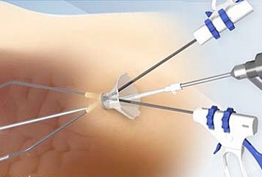
Technique of knee arthroscopy
Before carrying out arthroscopy, the so-called tourniquet is applied to the thigh of the patient, which helps to reduce the degree of bleeding in the joint cavity. This improves the visibility of the doctor who conducts arthroscopy. For carrying out arthroscopy in the knee joint area, three small (5 - 7 mm) skin incisions are performed. Through these incisions, joints are inserted into the joint cavity. One of these tools has a light source and a video camera lens on the end. Information from it is transmitted to the monitor screen. Through another tool, which is a hollow tube, a sterile transparent liquid is inserted into the half of the knee joint. This allows you to increase the volume of the joint cavity, which facilitates the inspection and therapeutic manipulations. Through the third incision, the main working tool is inserted into the joint, with the help of which medical manipulations are performed (removal of the meniscus, restoration of the ligaments, etc.). After the arthroscopy, the instruments are removed, the injected liquid is pumped out and, if necessary, a solution with a drug is added (antibiotics, anti-inflammatory drugs). Sterile bandages are applied to the incisions and a pressure bandage is applied to the knee area.
After arthroscopy
Usually it takes 1 to 2 days to stay in the hospital. Anesthetics are prescribed. For 2 - 3 days, a plaster lingeta is applied to create a temporary rest in the joint. From the first day the patient is recommended to train the periarticular muscles to prevent their atrophy. A week later you can completely bend the knee joint. On the 5th - 6th day, a gradual load on the joint is allowed.


Healthy knee joint Joint, affected by deforming osteoarthritis
A special place is occupied by arthroscopy in the treatment of deforming osteoarthritis of the knee joints, allowing to prolong the "service life" of the joint, and to postpone the time of more serious and serious operations (corrective osteotomy, total endoprosthetics).
COMPLICATIONS OF ARTHROSCOPY
Like any other surgery for arthroscopy, there may be, although rare, some complications.
These include:
- Complications caused by anesthesia are not associated with arthroscopic surgery, as such.
- Complications from the vascular system: an extremely rare complication in which a popliteal artery or vein is touched during surgery.
- Complications from the nervous system: they are manifested by the appearance of a zone of "anesthesia" or sensation of goosebumps in the joint region and are associated with damage to the branches of the nerves. Over time, these manifestations disappear.
- Stretching of the inner lateral ligament: can be obtained during enhanced manipulations aimed at increasing the distance between the femur and the tibia in the study of menisci
- Thromboembolic complications: very rare.
- Arthritis - is associated with getting into the joint of an infection.
- Hemarthrosis: this is the appearance of a severe and painful hemorrhage in the joint. It is a rare complication after arthroscopy.
- The swelling of the articular fluid often appears when the activity is resumed too quickly after the operation.
Recommendations for patients after arthroscopic knee surgery
Patients who underwent arthroscopic surgery in the first weeks are recommended:
- Walk with full support on the leg.
- Relieve the venous outflow by lifting your leg.
- Thigh muscle training
- It is not recommended to squat or bend the knee more than 90 degrees.